I. Spectral absorption functions of pigments.
Light is perceived, or detected, when photons of light are captured by pigments in photoreceptor cells (rods and cones). The capture of photons by the pigments results in an electrical stimulus which ultimately is transmitted to the brain. In general the more photons that are captured in a short amount of time, the stronger the stimulus to the brain. Photons which travel into the rods and cones are not always absorbed by the pigment. Some percentage will travel right on through and cannot be detected. Each photon which strikes the retina has a certain probability (i.e. likelihood) of being captured.
Photons come in different wavelengths (or frequencies). It turns out that the probability that a given pigment will absorb a photon of a given wavelength depends on the wavelength. If 1000 photons per second of some wavelength strike the retina, but the probability of each being captured is only 50%, then on average only 500 photons per second will be captured. Since the number of photons captured by the pigment determines how bright the stimulus appears, the brightness of a given light stimulus will depend both on (1) the intensity, or number of photons per second and (2) on the wavelength of the photons, since capture probability depends on wavelength.
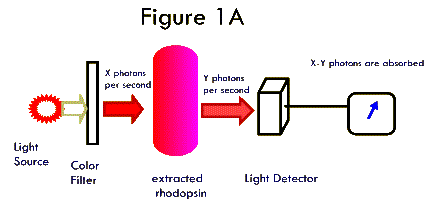
It is possible to extract large quantities of the pigment rhodopsin (the pigment found in rods) from the eyes of nocturnal animals. We can take a bottle of rhodopsin and shine small lights of different wavelengths through it (Figure 1A). We can measure the intensity (photons per second) of the light striking the pigment, and then measure the intensity of the light which has passed through the pigment. The difference in these two values represents the amount of light absorbed. We can repeat these measurements of light of the same intensity at a number of different wavelengths. If we then plot the data -- percent light absorbed versus wavelength -- we have a measure of the likelihood that a photon of any given wavelength will be absorbed by the rhodopsin. This curve (Figure 1B) is called the spectral absorption function of rhodopsin.
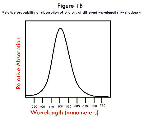
This function tells us how well rhodopsin absorbs light of different wavelengths. It turns out that it also predicts very nicely how bright light of different wavelengths will look to an observer using his/her rods to view stimuli. In other words, a stimulus of 50 photons per second at 510 nm will look roughly twice as bright as a stimulus of 50 photons per second at 560 nm. So, the brightness depends to a large degree on its wavelength!
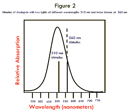
Recall our exercise in lab. We determined that rod-based vision does not give us information about color. This is a little puzzling since Fig. 1B shows that different colors stimulate rods to differing degrees. However, consider the following (illustrated in Fig. 2). Suppose we stimulate a rod with light at 560 nm and a stimulus intensity of 500 photons per second. Now lets say we stimulate the rod with light at 510 nm at an intensity of 500 photons per second. The second stimulus will look twice as bright as the first. Now suppose we go back to the 560 nm stimulus and increase the stimulus intensity to 1000 photons per second. The response of the rod will be twice what it was to 500 photons per second. However this response will be IDENTICAL to the response to the 510 nm stimulus at 500 photons per second. In other words there is an inherent ambiguity in how rods respond. If two stimuli appear to be different in brightness, it could be either because (1) they differ in wavelength, or (2) because they differ in stimulus strength. This is why we cannot distinguish colors with our rods. We have no way of telling whether we are seeing stimuli that differ in intensity or differ in wavelength.
II. Spectral absorption functions of cones
The pigment found in cones is very similar to that found in rods, except that (1) there is a lot less of it, and (2) the different cones possess pigments which differ in their absorption functions. It is possible to extract individual cones from the retina and pass tiny light through their outer segments (where the pigment is found) and measure the relative absorption of different wavelengths of light. This process is called microspectrophotometry. Doing this it has been found that humans (and most monkeys in which most of this work was actually carried out) possess three types of cones. The absorption functions of the pigments in each of the three cone types are shown in Figure 3. Note that the wavelength that is absorbed best is different for each type of cone. This value -- the wavelength of peak asorption -- is called lmax. (read "lamda max"). The three classes of cones in the human retina are referred to as the S-cone (short wavelength), M-cone (medium wavelength), and L-cone (long wavelength).
Just as with rods, the response of an individual cone is directly related to (1) the intensity of the stimulus falling on it (in photons per second) and (2) the wavelength of the stimulus.
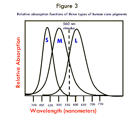
Suppose we stimulate the cones with a single wavelength of light = 560 nm. at an intensity of 1000 photons per second. Note that this stimulus gives equal stimulation to the M-cone and the L-cone. A stimulus of this type appears yellow to most human observers. Suppose now we double the intensity of the stimulus to 2000 photons per second. The M-cone will respond twice as strongly. The L-cone will respond twice as strongly. However, the ratio of response of the two cones will not change!
THIS IS THE ESSENCE OF COLOR VISION. THE BRAIN RECORDS THE RATIO OF RESPONSES FROM THE DIFFERENT CLASSES OF CONES.
III. Metamers
So far we have only talked about so-called spectral colors, those that consist of only a single wavelength. In the real world many objects reflect a whole range of different wavelengths. Similarly many light sources (the sun, for example) produce a whole range of wavelengths. How are these colors perceived?
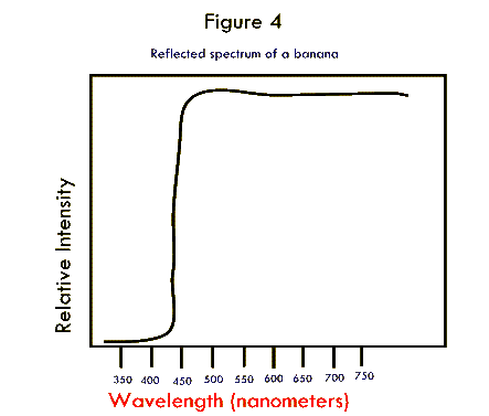
A stimulus color that consists of many different wavelengths, with different intensities at each wavelength is called a light spectrum, or spectral pattern. This could represent the spectrum of a light source, or the light reflected from some surface. Consider the spectral pattern shown in Fig. 4. This plot shows light that might reflect off of a banana illuminated by sunlight. Note that wavelengths shorter than 500 nm are absorbed by the banana, but all wavelengths longer than 500 are reflected. WHY DOES THIS LOOK YELLOW?
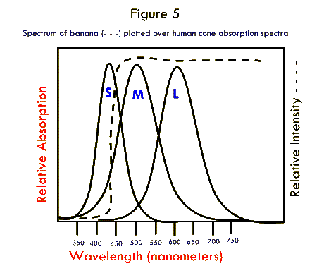
In Figure 5 the same spectrum is shown plotted on top of the spectrum of the 3 classes of human cones. The total stimulation of each cone is equivalent to the number of photons absorbed. To calculate the relative number of photons absorbed we multiply the stimulus spectrum x the absorption spectrum or measure the area of overlap between the two curves.
If we do this we find that this stimulus hits the L cone and M cone just about equally. In other words the ratio of output from the two cones is 1:1! This is the same as it was for the spectrally pure stimulus at 560 nm. ANY STIMULUS THAT GIVES APPROXIMATELY EQUAL STIMULATION TO THE L AND M CONE APPEARS YELLOW.
Two colors that have very different spectra, but that are perceived as being identical are called metamers.
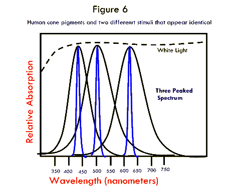
Look at the two stimulus spectra shown in Figure 6. The dotted line shows the spectrum from a white light. The solid line shows a spectrum consisting of three sharp peaks. These two spectra are metamers! They look identical. That is because we perceive white when there is roughly equal stimulation of all 3 of our cone classes.
IV. General rules for perceived colors
In general stimuli that primarily stimulate the S-cone look blue, those that stimulate the M-cone more than either S or L look green. Those that stimulate M and L cones tend to look yellow. Stimuli that primarily stimulate the L cone appear read. Different combinations of stimulation give all the different shades of color. Spectra which mainly stimulate the S and the L-cone together look purple. This is a unique sensation because there is no single spectral color that can cause the same sensation.
In general the perception of color is based on the ratio of stimulation of the three classes of cones. There is an interesting consequence of this fact. One can change the amount of stimulation of any one cone by changing the intensity of the light striking it. This means that if one shines 3 different lights on the eye, and each of the lights primarily stimulates a different cone, one can create EVERY POSSIBLE COLOR SENSATION BY ALTERING THE RATIO OF STIMULATION OF THE THREE CONES. THIS CAN BE DONE BY CHANGING THE INTENSITY OF STIMULATION FROM THE THREE LAMPS.
Last Modified: Tuesday, 04-Nov-2003 fleishml@union.edu